

01.03.2024
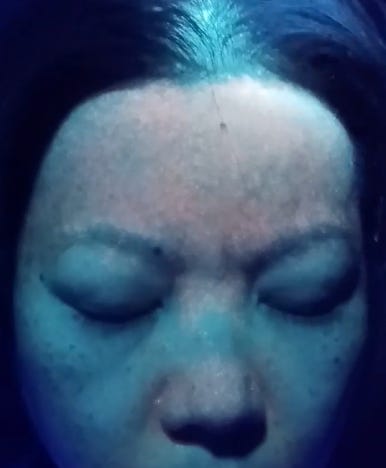
The recent findings of a facial glow in the faces of the C19 injected has shocked many people, while others are quick to dismiss or ignore the findings that indicate nanotechnology contamination and synthetic biology modification.

Image Courtesy: Justin Coy, PhD, Individual with 1 Moderna C19 Injection and 3 PCR tests shows orange flourescent skin tattoo. In the white sclera are 2 flourscent bright spots on boht sides of the iris consistent with Eye of Horus Phenomenon In this follow up substack, I am posting the images that Justin Coy, PhD was talking about regarding the Eye of.., Read full story
But is this finding really so unusual given what the biotechnology sector has been working on for the last two decades? Did you know of the obsession of synthetic biology to introduce florescent proteins via SV40 plasmids in the the DNA of just about everything living?
Is there any correlation with C19 injected glowing orange and the SV40 plasmids that were found in the C19 vials?
Here is how fluorescent jellyfish protein sequences get inserted via SV40 promoters into a host DNA:
Introduction to Fluorescent Proteins
Fluorescent Protein Vectors and Gene Transfer
Fluorescent proteins are quite versatile and have been successfully employed in almost every biological discipline from microbiology to systems physiology. These ubiquitous probes have been extremely useful as reporters for gene expression studies in cultured cells and tissues, as well as living animals. In live cells, fluorescent proteins are most commonly employed to track the localization and dynamics of proteins, organelles, and other cellular compartments. A variety of techniques have been developed to construct fluorescent protein fusion products and enhance their expression in mammalian and other systems.The primary vehicles for introducing fluorescent protein chimeric gene sequences into cells are genetically engineered bacterial plasmids and viral vectors.
Fluorescent protein gene fusion products can be introduced into mammalian and other cells using the appropriate vector (usually a plasmid or virus) either transiently or stably. In transient, or temporary, gene transfer experiments (often referred to as transient transfection), plasmid or viral DNA introduced into the host organism does not necessarily integrate into the chromosomes, but can be expressed in the cytoplasm for a short period of time. Expression of gene fusion products, easily monitored by the observation of fluorescence emission using a filter set compatible with the fluorescent protein, usually takes place over a period of several hours after transfection and continues for 72 to 96 hours after introduction of plasmid DNA into mammalian cells. In many cases, the plasmid DNA can be incorporated into the genome in a permanent state to form stably transformed cell lines. The choice of transient or stable transfection depends upon the target objectives of the investigation.

The basic plasmid vector configuration useful in fluorescent protein gene transfer experiments has several requisite components. The plasmid must contain prokaryotic nucleotide sequences coding for a bacterial replication origin for DNA and an antibiotic resistance gene. These elements, often termed shuttle sequences, allow propagation and selection of the plasmid within a bacterial host to generate sufficient quantities of the vector for mammalian transfections. In addition, the plasmid must contain one or more eukaryotic genetic elements that control the initiation of messenger RNA transcription, a mammalian polyadenylation signal, an intron (optional), and a gene for co-selection in mammalian cells. Transcription elements are necessary for the mammalian host to express the gene fusion product of interest, and the selection gene is usually an antibiotic that bestows resistance to cells containing the plasmid. These general features vary according to plasmid design, and many vectors have a wide spectrum of additional components suited for particular applications.
Illustrated in Figure 7 is the restriction enzyme and genetic map of a commercially available (BD Biosciences Clontech) bacterial plasmid derivative containing the coding sequence for enhanced yellow fluorescent protein fused to the endoplasmic reticulum targeting sequence of calreticulin (a resident protein). Expression of this gene product in susceptible mammalian cells yields a chimeric peptide containing EYFP localized to the endoplasmic reticulum membrane network, designed specifically for fluorescent labeling of this organelle. The host vector is a derivative of the pUC high copy number (approximately 500) plasmid containing the bacterial replication origin, which makes it suitable for reproduction in specialized E. coli strains. The kanamycin antibiotic gene is readily expressed in bacteria and confers resistance to serve as a selectable marker.
Additional features of the EYFP vector presented above are a human cytomegalovirus (CMV) promoter to drive gene expression in transfected human and other mammalian cell lines, and an f1 bacteriophage replication origin for single-stranded DNA production. The vector backbone also contains a simian virus 40 (SV40) replication origin, which is active in mammalian cells that express the SV40 T-antigen. Selection of stable transfectants with the antibiotic G418 is enabled with a neomycin resistance cassette consisting of the SV40 early promoter, the neomycin resistance gene (aminoglycoside 3’-phosphotransferase), and polyadenylation signals from the herpes simplex virus thymidine kinase (HSV-TK) for messenger stability. Six unique restriction enzyme sites (see Figure 7) are present on the plasmid backbone, which increases the versatility of this plasmid.
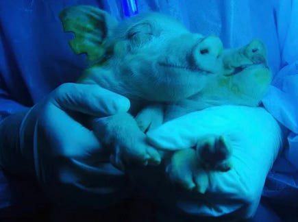
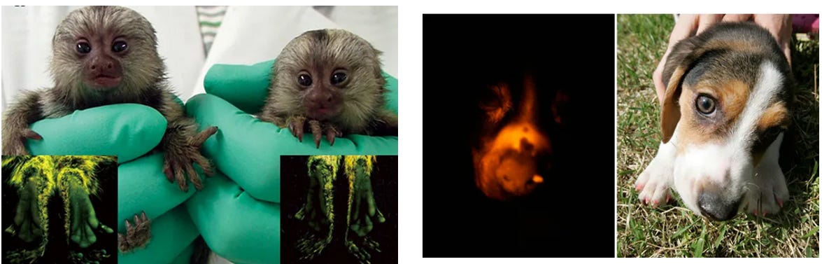
Have they done this successfully already? Yes, it started in animals almost 2 decades ago, animals that glow in different colors under blue light.
7 genetically modified animals that glow in the dark
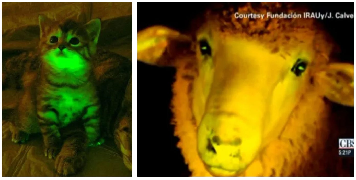
In 2008, scientists in Taiwan claimed to have a world first: Pigs that glowed from the inside out. While other researchers had bred partially fluorescent pigs, these genetically modified pigs had not only glowing skin and eyes, but also organs, including the heart. Scientists added DNA from fluorescent jellyfish to more than 260 pig embryos, which were then implanted into eight different sows, four of which became pregnant. The result was three male piglets whose eyes, teeth, and snouts had a slightly greenish tint during the day, but would glow entirely green in the dark after being introduced to a blue light.
Genetic engineered glowing dogs and marmosets Kei glow under UV light
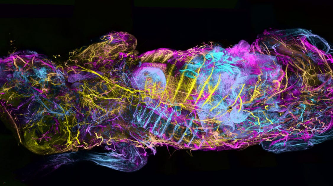
They have taken this even further now by completely mapping an organism with fluorescent protein expression.
How sick can these synthetic biology FREAKS in their blasphemy and disrespect against all good ethic and moral laws and being rights get?
STOP this SCIENCE!
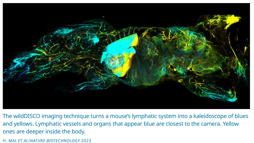
Fluorescent proteins light up the entire nervous system of a dead and transparent mouse, using a new technique employed here. The animal’s head is on the left. Colors show how deep nerve cells are in the animal, from blue (closest to the camera) to pink and then yellow (farthest away).
An improved technique starts by chemically removing cholesterol from the tissues of dead mice. That cholesterol is an essential part of cell membranes. Taking it out created spongelike holes in tissues — without destroying them. Those holes allowed better use of chemicals that can color, or label, structures of interest.
Targeted antibodies can move through the holes to reach every corner of the body. They bind to proteins of interest everywhere they reach. Under fluorescent light, they can make targeted parts of the body glow.
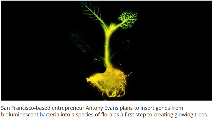
The next generation of glowing plants Using nanoparticles that store and gradually release light, engineers create light-emitting plants that can be charged repeatedly.
Using specialized nanoparticles embedded in plant leaves, MIT engineers have created a light-emitting plant that can be charged by an LED. After 10 seconds of charging, plants glow brightly for several minutes, and they can be recharged repeatedly.
These plants can produce light that is 10 times brighter than the first generation of glowing plants that the research group reported in 2017.
“We wanted to create a light-emitting plant with particles that will absorb light, store some of it, and emit it gradually,” says Michael Strano, the Carbon P. Dubbs Professor of Chemical Engineering at MIT and the senior author of the new study. “This is a big step toward plant-based lighting.”
This is an article from 2013 discussing inserting the Luciferin gene into plants.
Scientists genetically engineered the very first glow-in-the-dark plant in the 1980s, a tobacco plant with a firefly gene inserted into it. Historically, what has been the purpose of doing this?
The first time, I think, was just a demonstration project. But scientists have used it since to study things like root growth. They really use it for basic research purposes. Traditionally, what they’ve done is insert the gene for luciferase [an enzyme from a luminescent organism] along with a promoter [a region at the beginning of a gene that tells a cell to start transcription, the first step to producing a protein] and then add the luciferin [a chemical that produces light when oxidized] manually. They have even had these glowing plants up on the International Space Station, so it is a pretty well established technique.
What gene are you adding to create the glow?
We are using genes from Vibrio fischeri. It is marine bacteria.
How is this done? Can you take me through the process of creating a glowing plant?
We start with software called Genome Compiler. Genome Compiler allows us to search for gene sequences and then modify those gene sequences in a nice graphical user interface. We use that software to look up the Vibrio fischerigenes, and then we do something called code and optimization, which basically adjusts the sequences so that they [work] in plants instead of in bacteria. We then synthesize the DNA. There is a “print” button, and we “print” that DNA. That emails the file to a company, who makes the DNA for us. They FedEx that back to us, and then we do two things.
First, we insert the DNA into some bacteria called agrobacterium. That bacterium is very clever, it has figured out how to do genetic engineering on its own. [The bacterium] inserts the DNA into the female gametes of the plant. We can grow the seeds that come from those flowers, and we’ll have the DNA that we designed on the computer in the plant. The second thing we are doing is using a gene gun, which is a piece of equipment that fires the DNA at high velocity into the cells of the plant. Some of those cells will absorb the DNA and start to express it.
Are C19 injected humans just another synbio project in the globalist satanic technocratic transhumanist quest to genetically modify all of Gods creation? Luciferase? SV40? Really?
Its time to order a 365nm UV light flashlight and take a look for yourself.
Then get a live blood analysis done by someone who knows how to look for self assembly nanotechnology.
You answer the question for yourself.
What comes next?

Hinterlasse einen Kommentar